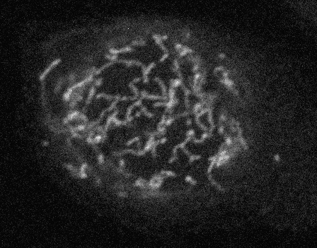
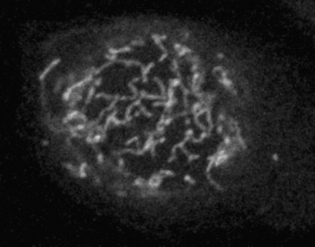
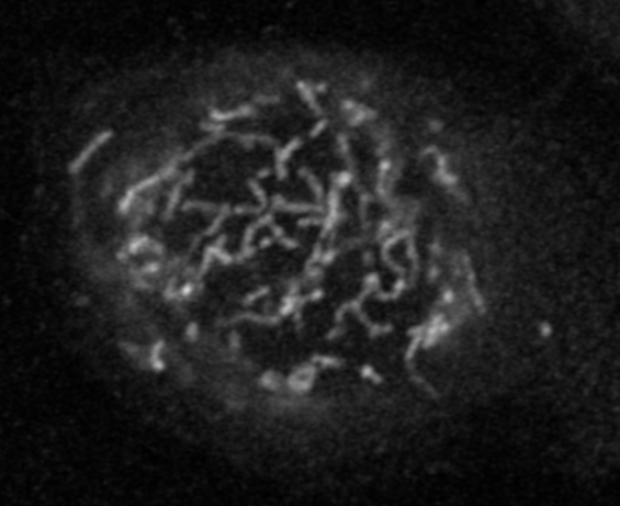
Mammalian cell culture labeled with fixable mitotracker red, fixed with fresh PFA, and images with a Zeiss LSM 800 with AiryScan.
For standard confocal, pinhole was set to 1 airy unit as defined by Zeiss with a 63X N.A. 1.4 planapochromat lens. The Airyscan light path was aligned to a perfect bright spot at the center of the detector with symmetric decay radiating outwards.
For this single optical section comparison, images were taken within 2 um of the coverslip.
Standard confocal pixel size is 0.063 um and the AiryScan pixel size is 0.049 um.
Order of images: confocal raw, confocal with median filter radius 1, Airyscan.
Order of images: confocal with median filter radius 1, Airyscan.
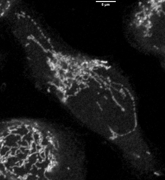
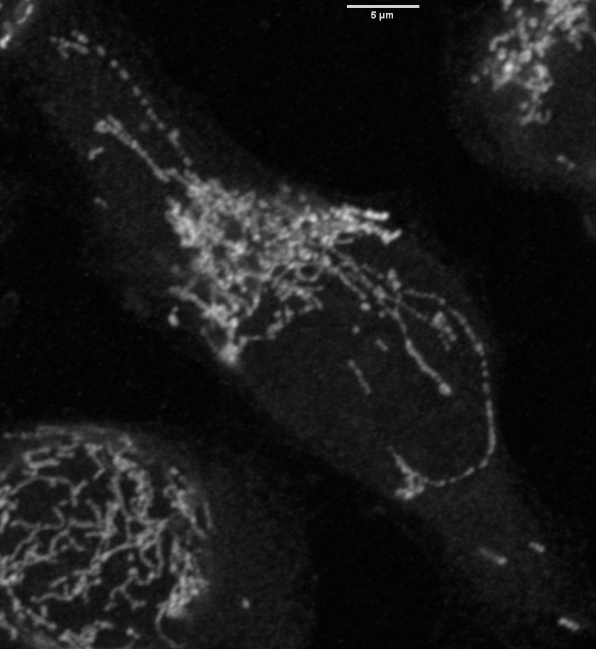
Projections shown at zoom 67%.
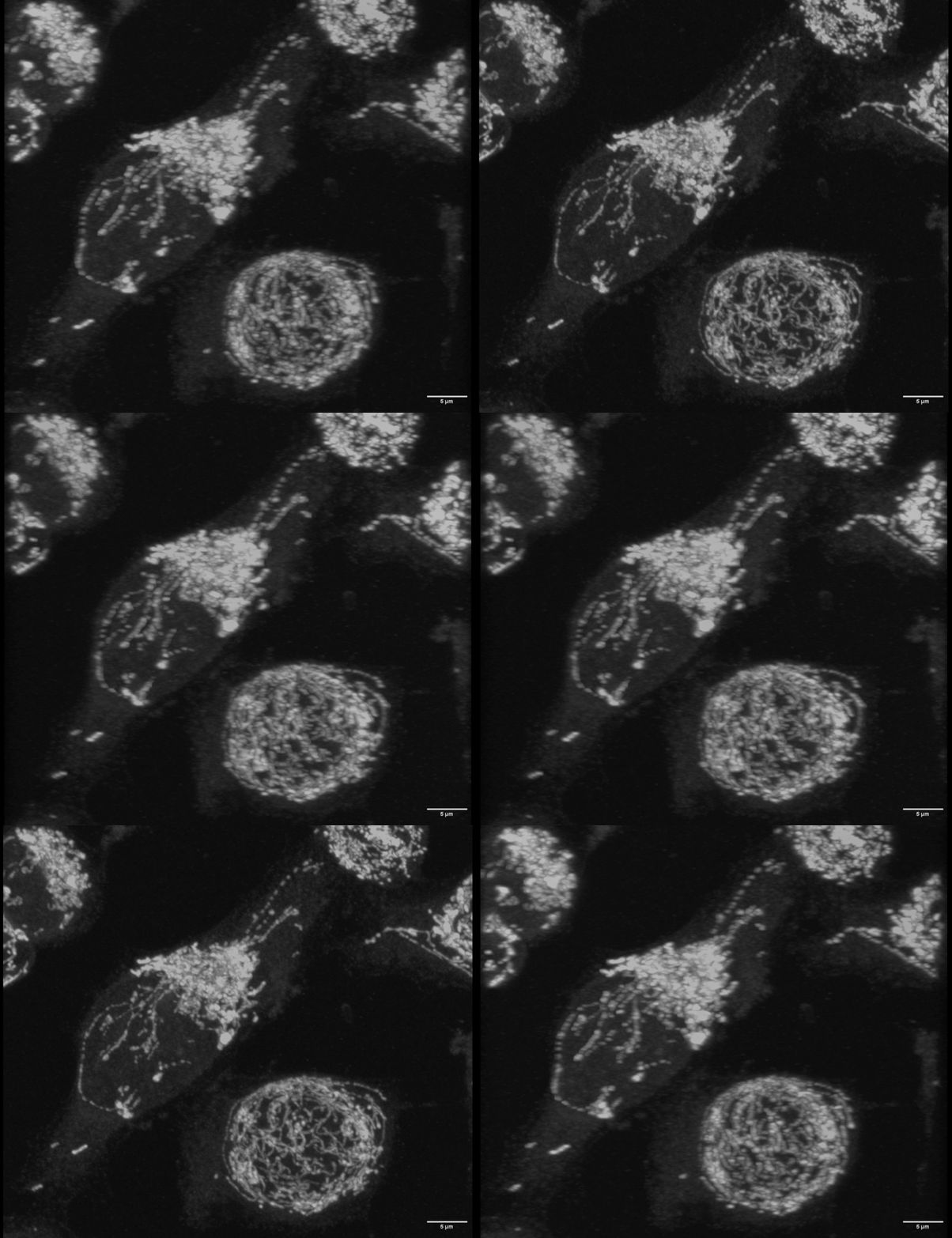
In this case, Airyscan and confocal comparison shows that with non diffraction limited objects, Airyscan does not provide significant benefits.
Due to the long lengths, curvature, and 3D nature of the mitochondria, could TEM capture these qualities? Could TEM capture more phenotypes, such as the apparent association with filaments (which light microscopy may be able to determine with counter staining)?
last updated 20261026